42 a well labelled diagram of a binocular microscope
Labeled Well Labelled Labeled Binocular Compound Microscope Diagram of a well labelled binocular microscope. Binocular microscope drawing at getdrawings com free for personal. An ocular lenses and objective lenses. Understanding the parts and features of a binocular microscope allows greater use of the microscope in the examination of specimens. Microscope- Definition, Parts, Functions, Types, Diagram, Uses It is a type of fluorescence microscope that is used to produce 2-D or 3-D images of relatively thick specimens. In this type, the excitation light is focused on a specific spot of sample lying on the focal plane. The focus spot is optically manipulated to scan the entire sample and generate a 3-D image.
A Study of the Microscope and its Functions With a Labeled Diagram The microscope is an important instrument in the world of biological science. Diagrams have always been of great help in understanding both the structural and functional aspects of entities. These labeled microscope diagrams and the functions of its various parts, attempt to simplify the microscope for you.

A well labelled diagram of a binocular microscope
Parts of Stereo Microscope (Dissecting microscope) - labeled diagram ... Labeled part diagram of a stereo microscope Major structural parts of a stereo microscope. There are three major structural parts of a stereo microscope. The viewing Head includes the upper part of the microscope, which houses the most critical optical components, including the eyepiece, objective lens, and light source of the microscope. 2.4 Parts of the Petrographic Microscope - Introduction to Petrology If available, the camera attached to the microscope will likely produce better-quality images. The Accessory Plates. Figure 2.4.17 shows examples of accessory plates including a quartz wedge, a 530 nm plate, and a 1/4 λ plate. These can be inserted into the microscope (Figure 2.4.2) above the objectives. The accessory plates are used to conduct advanced optical tests such as optical sign determination. Sperm Under Microscope with Labeled Diagram Let's make it clear (structure of Sertoli cell) from the below-mentioned labeled diagram. Here, the spermatogenic cells are adherent to the Sertoli cells. In the next part of this article, you will know and identify all of these spermatogenic cells from the seminiferous tubule along with the Sertoli cells.
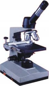
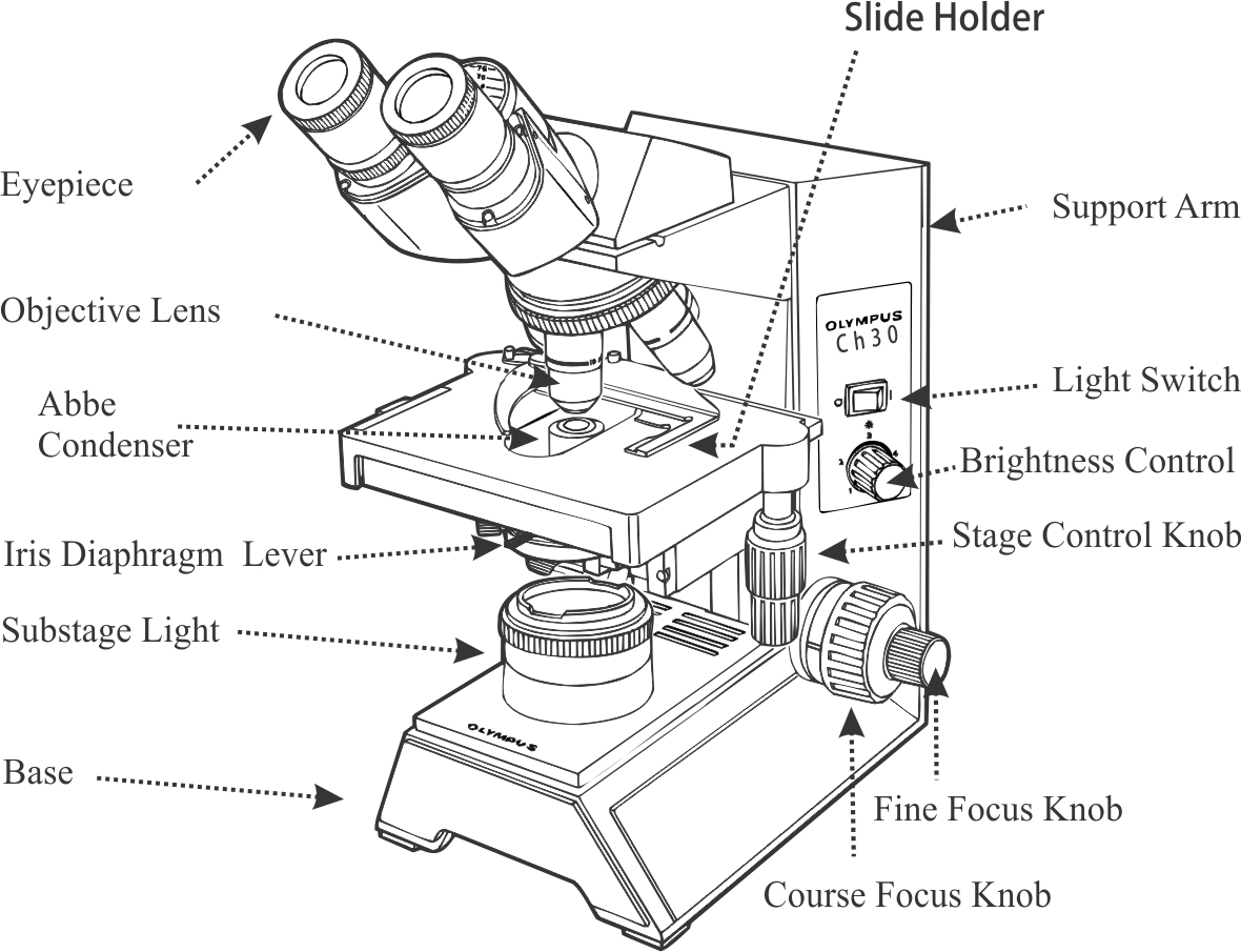
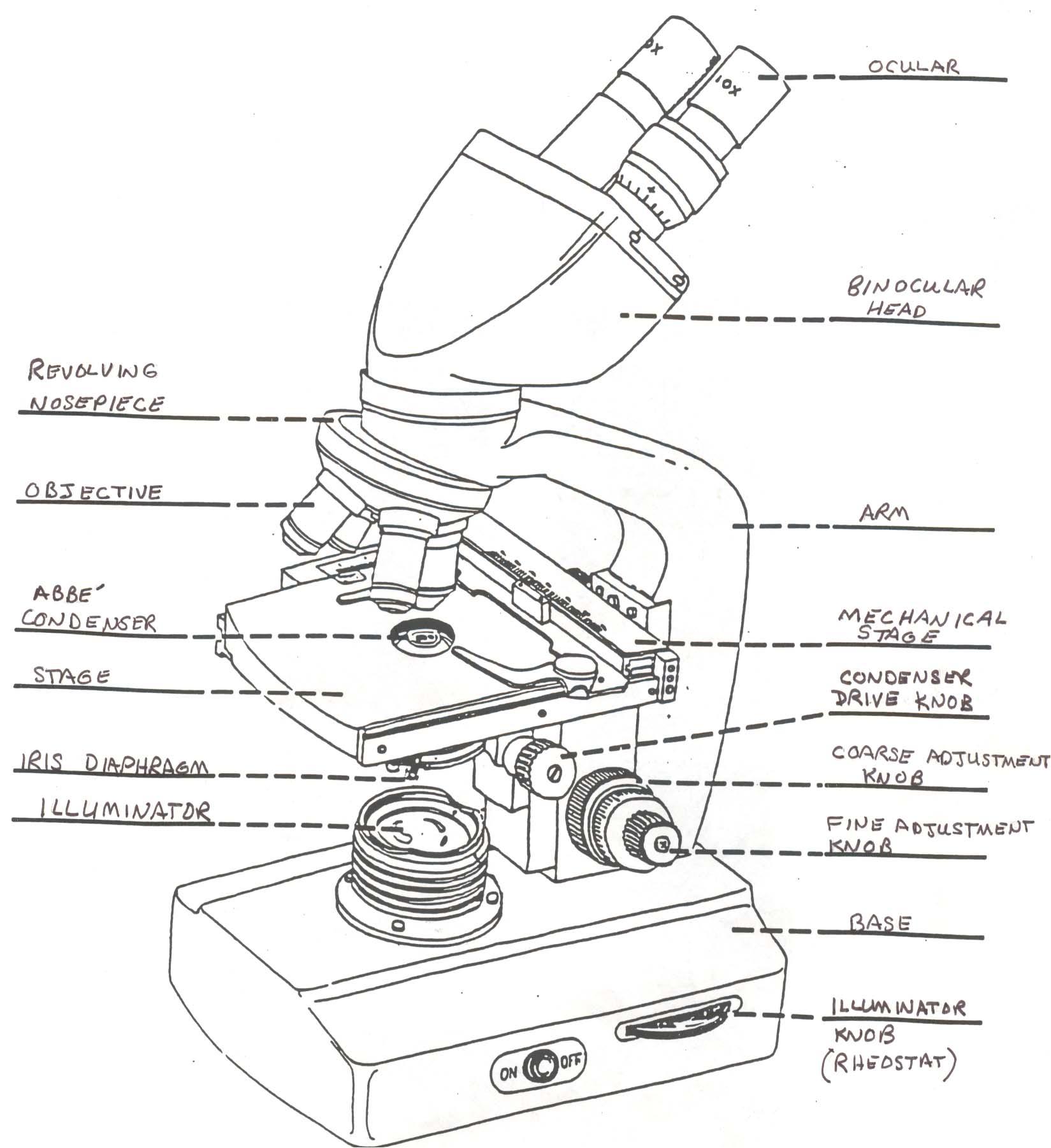
A well labelled diagram of a binocular microscope. Microscope Diagram Labelled - Micropedia Binocular Microscope Drawing At Getdrawings Free Download. Describe The Structure Of Compound Microscope With Well Labelled. Fluorescent Microscopy Lnf Wiki. Microscopy. ... 28 Label Microscope Diagram Lab 4 Biology 111 With Bishop. Microscopy. Microscope Labeled Keen Rsd7 Org. Parts of Binoculars and Their Functions Guide - TheOptics.org The objective lens captures the light from where the binoculars are pointed and reflects it onto the prisms. However, the image from the objective is usually inverted both horizontally and vertically. The first prisms in the binoculars would invert the image vertically while the other one would invert horizontally. What Are the Parts and Functions of a Binocular Microscope? Micah Elizabeth Scott/CC-BY-2.. The parts of a binocular microscope are the eye piece (ocular), mechanical stage, nose piece, objective lenses, condenser, lamp, microscope tube and prisms. Each part plays an important role in the microscope's function. Eye piece (ocular): The dual binocular eye piece contains the microscope's lenses and gives the ... Labelled Diagram of Compound Microscope - Biology Discussion The below mentioned article provides a labelled diagram of compound microscope. Part # 1. The Stand: The stand is made up of a heavy foot which carries a curved inclinable limb or arm bearing the body tube. The foot is generally horse shoe-shaped structure (Fig. 2) which rests on table top or any other surface on which the microscope in kept.
Light Microscope- Definition, Principle, Types, Parts, Labeled Diagram ... Brightfield Light Microscope (Compound light microscope) This is the most basic optical Microscope used in microbiology laboratories which produces a dark image against a bright background. Made up of two lenses, it is widely used to view plant and animal cell organelles including some parasites such as Paramecium after staining with basic stains. Microscope Labeling Diagram | Quizlet Unit 2 Lesson 5 - Punnett Squares and Pedigrees. 4 terms. PGFry210. Unit 2 Lesson 4 - Heredity. 9 terms. PGFry210. Upgrade to remove ads. Only $2.99/month. Compound Microscope Parts - Labeled Diagram and their Functions - Rs ... Many optical parts of a microscope work together to magnify and produce an image of the specimen placed on a slide. These parts include: Eyepiece. The eyepiece (or ocular lens) is the lens part at the top of a microscope that the viewer looks through. The standard eyepiece has a magnification of 10x. Label the microscope - Science Learning Hub Use this interactive to identify and label the main parts of a microscope. Drag and drop the text labels onto the microscope diagram. eye piece lens coarse focus adjustment high-power objective diaphragm or iris base fine focus adjustment light source stage Download Exercise Tweet
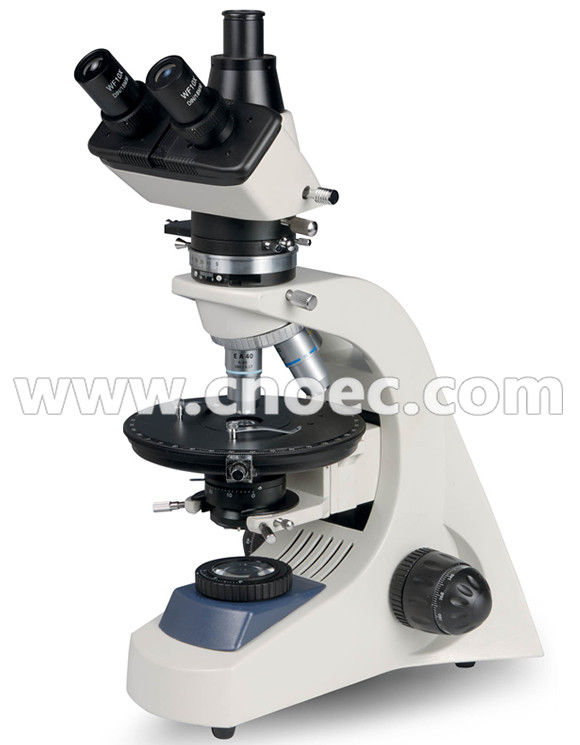
Microscope Quiz: How Much You Know About Microscope Parts ... - ProProfs Holds the slide in place. It is used to support the microscope when carried. 2. Magnification ranges from 10x to 40x. Holds the slide in place. Moves the stage up and down for focusing. 3. Moves the stage slightly to sharpen the image. Holds the high and low power objectives. PDF Binocular Microscope Sketch Diagram IT WITH THE BINOCULAR' 'Labeled Light Microscope Diagram Engine Diagram And 1 / 7. April 23rd, 2018 - Labeled Light Microscope Diagram Also Types Of Microscopes Furthermore Transmission Electron Microscopy Moreover 2007 08 01 Archive Also Paginas Para Colorir Microscopio I16110 As Well As Transmission Electron Microscopy Along With Post ... Compound Microscope Parts, Diagram Definition, Application, Working ... Compound Microscope is classified in two categories; A. Light Microscope. Light Microscope is further classified into four categories such as; Bright-field Microscope; Dark-Field Microscope. Phase-contrast Microscope. Fluorescent Microscope. B. Electron Microscope. Electron Microscope is further classified into three categories such as; Scanning Microscope Binocular Microscope Anatomy - Parts and Functions with a Labeled Diagram Microscope anatomy labeled diagram. Now, I will show you the different parts of the different types of microscopes (like the binocular, monocular, trinocular, electron microscope, confocal microscope, and others) with the labeled diagrams. First, let's see the different parts of the monocular or trinocular microscopes.
Tips for Buying a Light Microscope - Compound, Inverted and Stereoscope ...
Labelled Diagram Of A Light Microscope | Products & Suppliers ... Optical and Magnetic Tweezers. Advantages. Direct optical access. Piezo nanopositioner ready. High stability microscope with precision alignment. Fixed or automated objective lens positioning. Flexible design with multiple configurations. RM21 TM. The RM21 TM is a versatile microscope suitable for a variety of advanced microscopy and nanoscopy methods. The RM21 TM is available in 4 standard...
![Untitled Document [www.bio.utexas.edu]](http://www.bio.utexas.edu/courses/bio206/images/microscope labeled500pix.jpg)
Untitled Document [www.bio.utexas.edu]
Bacteria Diagram Unlabeled - celljail.blogspot.com Well labelled Diagram of Prokaryotic cell For board and NEET exams Bacterial cell. Bacteria diagram additionally indicates Periplasmic space thats a cellular compartment found in simple terms in bacteria which have an outer membrane and a plasma membrane. ... Blank Microscope Diagram Optics Binoculars Wiring Schematic. Cell diagram unlabeled ...

Amazon.com: Binocular Compound Microscope 40X-1600X Magnification ...
Parts of the Microscope with Labeling (also Free Printouts) 5. Knobs (fine and coarse) By adjusting the knob, you can adjust the focus of the microscope. The majority of the microscope models today have the knobs mounted on the same part of the device. Image 5: The circled parts of the microscope are the fine and coarse adjustment knobs. Picture Source: bp.blogspot.com.

Microscope - The Gemology Project
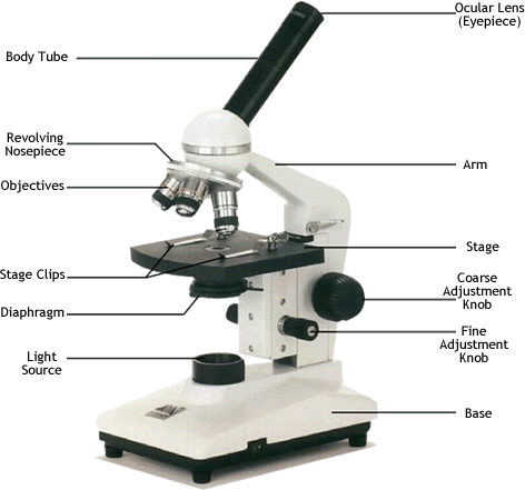
Microscope: Types of Microscope, Parts, Uses, Diagram - Embibe There microscope anatomy includes three structural parts, i.e. head, base, and arm. Head - This is also known as the body; it carries the optical parts in the upper part of the microscope.. Base - It acts as microscopes support.It also carries microscopic illuminators. Arms - The microscope arm connects the base and the head and the eyepiece tube to the microscope base.
What is the difference between monocular and binocular microscope? - Quora
Microscope Parts and Functions With Labeled Diagram and Functions How ... Body tube (Head): The body tube connects the eyepiece to the objective lenses. Arm: The arm connects the body tube to the base of the microscope. Coarse adjustment: Brings the specimen into general focus. Fine adjustment: Fine tunes the focus and increases the detail of the specimen.
MICROSCOPES Manufacturer, Supplier, Exporter
Labeling the Parts of the Microscope | Microscope World Resources Labeling the Parts of the Microscope. This activity has been designed for use in homes and schools. Each microscope layout (both blank and the version with answers) are available as PDF downloads. You can view a more in-depth review of each part of the microscope here. Download the Label the Parts of the Microscope PDF printable version here.
what is the highest magnification of a light microscope ...
Labeled Diagram Of A Stereo Microscope - GlobalSpec Climate diagrams of monthly mean climate conditions for the four sites studied are shown in Fig. 2. The cores were then labeled , fixed on wooden supports, air-dried and sanded with progressively finer grades of sandpaper (80, 180, 300, 500 grit) until the tree-ring structure was clearly visible under the stereo microscope .
Microscope Lenses Diagram - Micropedia
Compound Microscope: Definition, Diagram, Parts, Uses, Working ... - BYJUS Compound microscope is a type of optical microscope that is used for obtaining a high-resolution image. There are more than two lenses in a compound microscope. Learn about the working principle, parts and uses of a compound microscope along with a labeled diagram here.
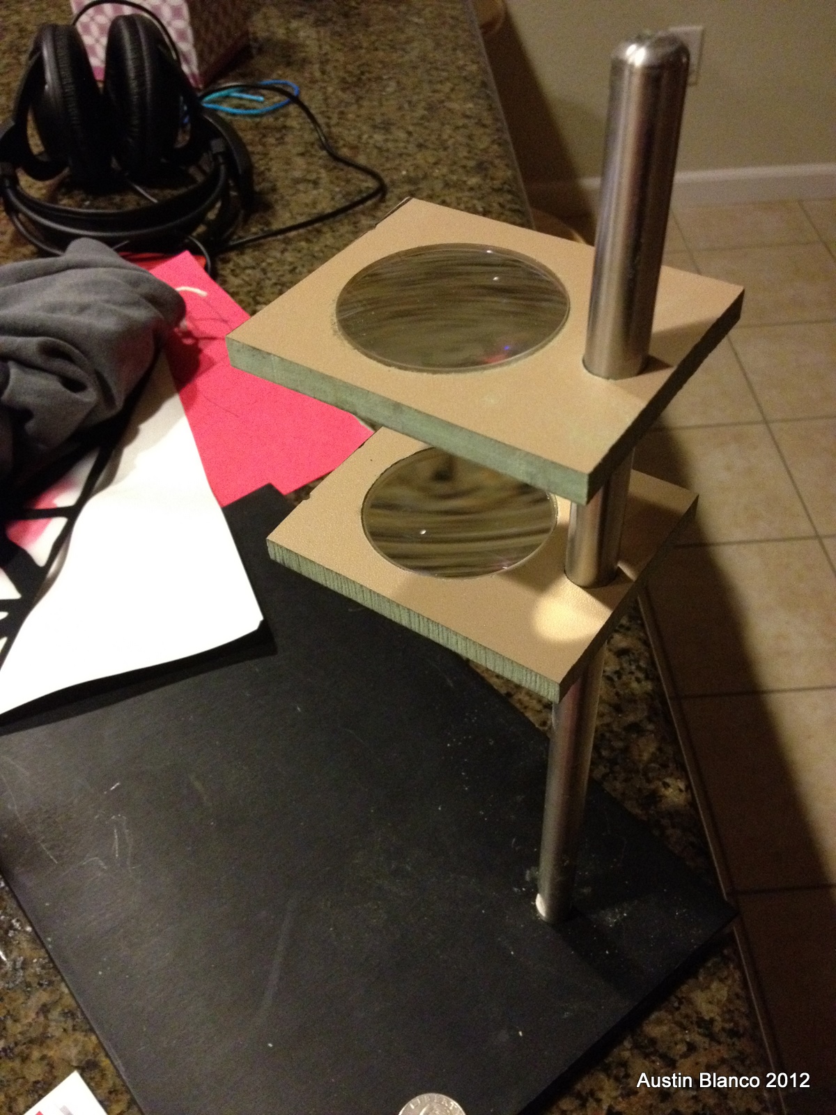
Making a microscope from binoculars - Austins Imaging Blog
What Are The Different Parts of a Binocular? - The Ultimate Guide!! b. Prism ( Porro & Roof Prism Type) -Corrects the inverted Image through the lenses to the original position of the object. c. Eyepiece/Ocular Lens - Presents the final image to the eyes. d. Casing/Binoculars Barrel - Provides a covering and fits the different binocular parts together. e.

Large binocular microscope | CURIOUS SCIENCE
Parts of a microscope with functions and labeled diagram Figure: Diagram of parts of a microscope. There are three structural parts of the microscope i.e. head, base, and arm. Head - This is also known as the body. It carries the optical parts in the upper part of the microscope. Base - It acts as microscopes support. It also carries microscopic illuminators.

A Well Labelled Diagram Of A Light Microscope - Top Label Maker
Sperm Under Microscope with Labeled Diagram Let's make it clear (structure of Sertoli cell) from the below-mentioned labeled diagram. Here, the spermatogenic cells are adherent to the Sertoli cells. In the next part of this article, you will know and identify all of these spermatogenic cells from the seminiferous tubule along with the Sertoli cells.

The Journal of Nadirsky.: Parts of Microscope (biology lesson)
2.4 Parts of the Petrographic Microscope - Introduction to Petrology If available, the camera attached to the microscope will likely produce better-quality images. The Accessory Plates. Figure 2.4.17 shows examples of accessory plates including a quartz wedge, a 530 nm plate, and a 1/4 λ plate. These can be inserted into the microscope (Figure 2.4.2) above the objectives. The accessory plates are used to conduct advanced optical tests such as optical sign determination.

Microscope Self-Test
Parts of Stereo Microscope (Dissecting microscope) - labeled diagram ... Labeled part diagram of a stereo microscope Major structural parts of a stereo microscope. There are three major structural parts of a stereo microscope. The viewing Head includes the upper part of the microscope, which houses the most critical optical components, including the eyepiece, objective lens, and light source of the microscope.
Microscope Drawing Template at PaintingValley.com | Explore collection ...
How to Use the Microscope
Microscope Drawing Template at GetDrawings | Free download
Post a Comment for "42 a well labelled diagram of a binocular microscope"