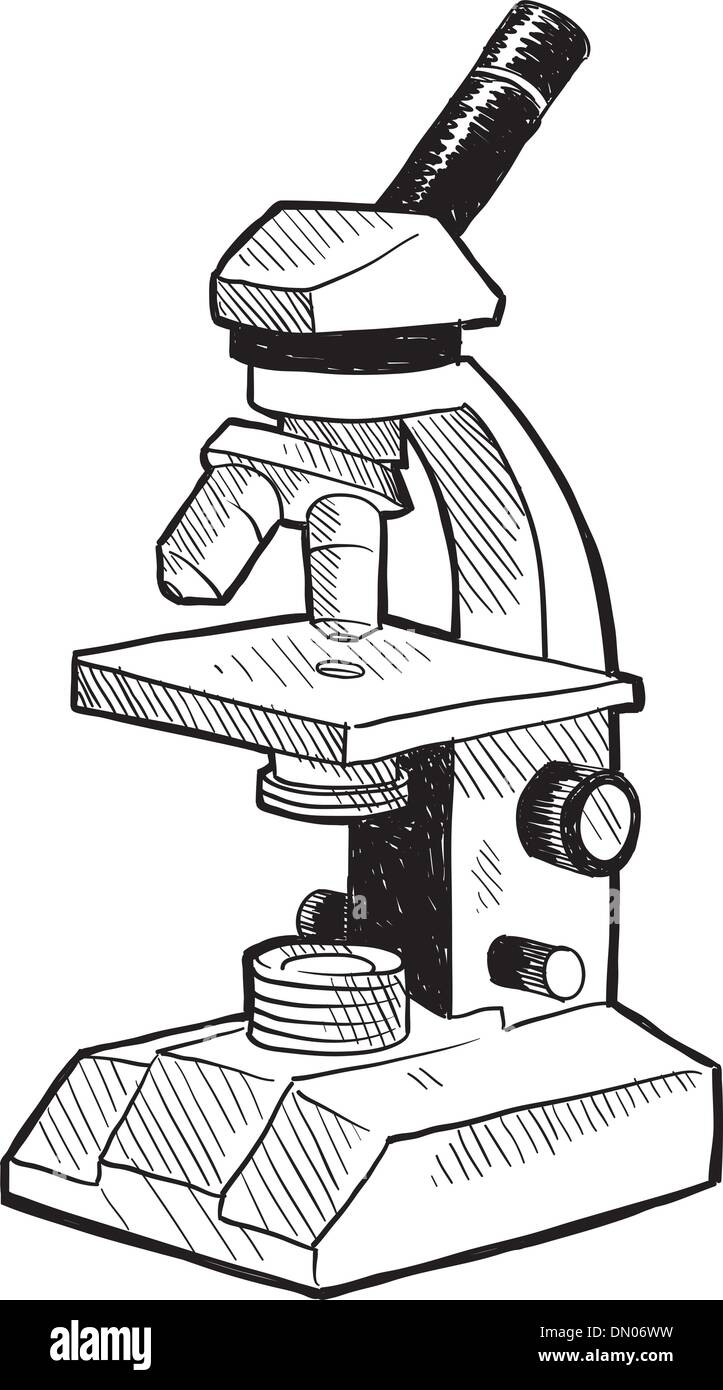
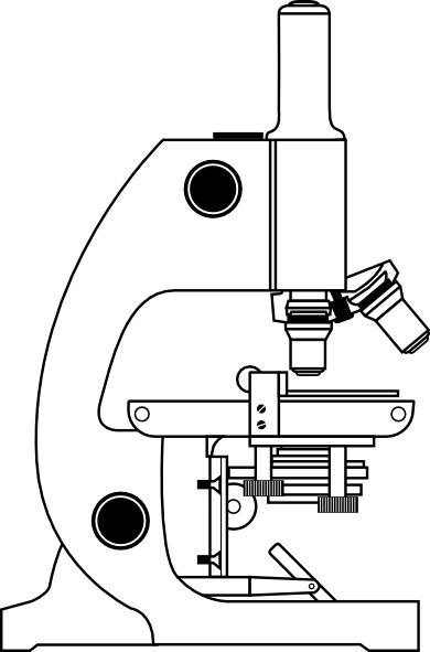
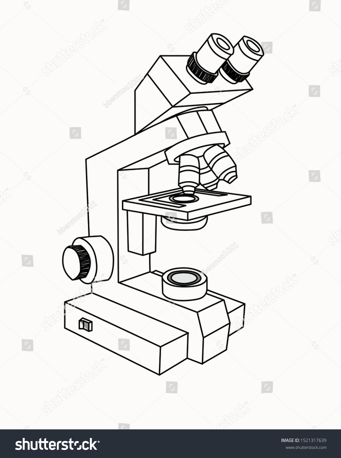
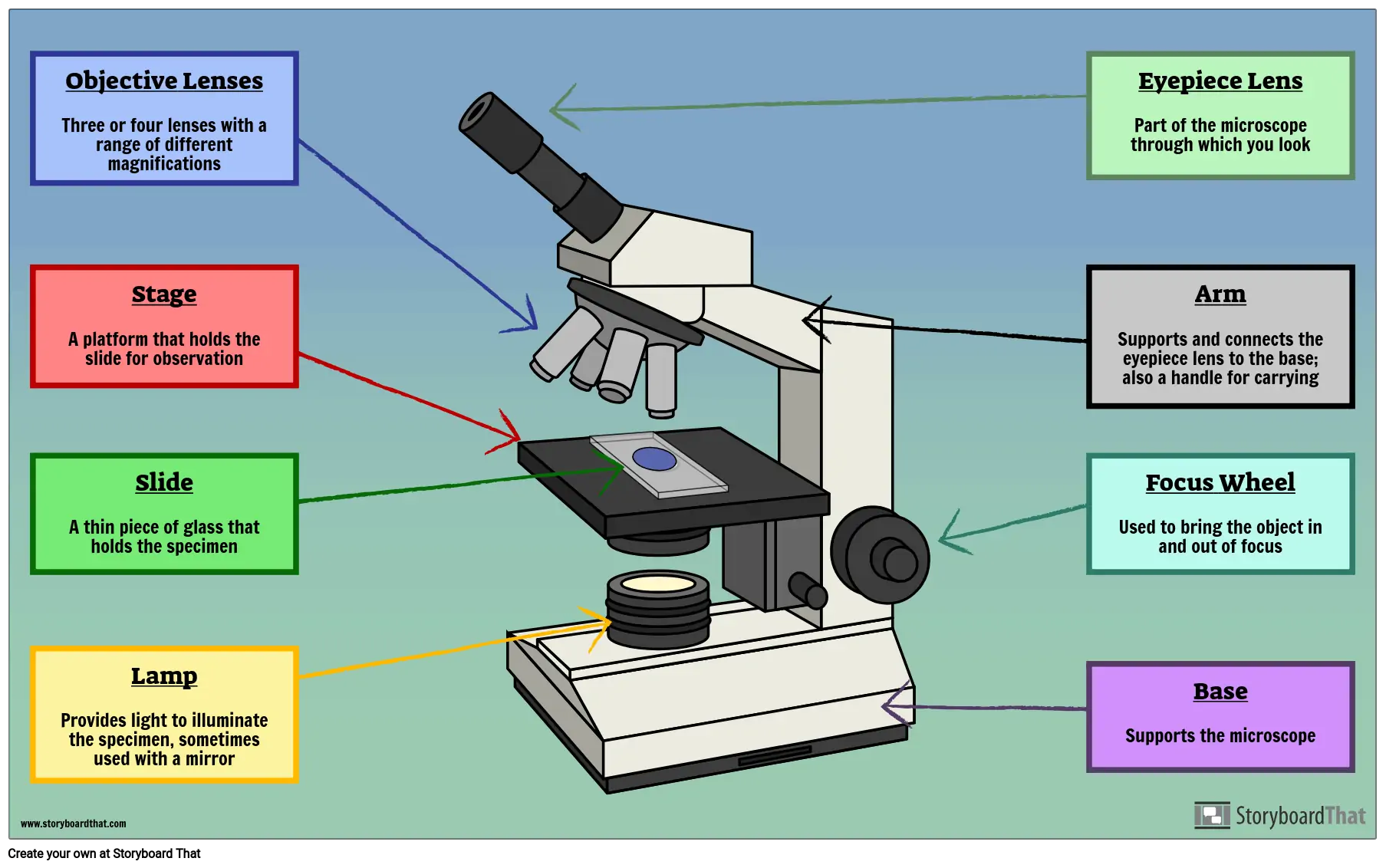
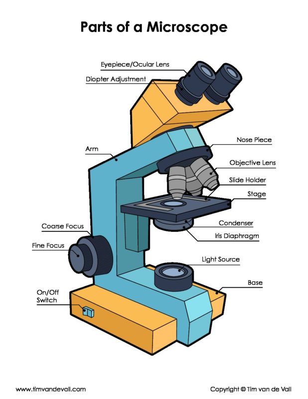
39 microscope drawing labeled
5 White Blood Cells Types and Their Functions - New Health Advisor Agranulocytes are free of visible grains under the microscope and include lymphocytes and monocytes. Together, they coordinate with one another to fight off things like cancer, cellular damage, and infectious diseases. Below, detailed information about each type will be discussed. 1. Neutrophils ECLIPSE Ti2 Series | Inverted Microscopes | Products | Nikon ... Leading platform for advanced imaging. The ECLIPSE Ti2 inverted microscope delivers an unparalleled 25mm field of view (FOV) that revolutionizes the way you see. With this incredible FOV, the Ti2 maximizes the sensor area of large-format CMOS cameras without making compromises, and significantly improves data throughput.
5 Steps of Gram Staining Procedure: How to Interpret the Results 2. Label the Slides Draw a circle under the slides using a marking pen designed for glassware. This will help to designate which area to prepare the smear in the following step. You can also label them with the organism's initials at the edge of each slide. Take care that the labels do not get in contact with the reagentsused forstaining. 3.

Microscope drawing labeled
STEM Tuesday - Fungi - In the Classroom - From The Mixed Up Files First prepare some microscope slides for students. Use a variety of fresh mushrooms, cut off the stems, and place the mushrooms gill-side down on top of the slide plates overnight. Make sure to label each slide. ... Students can draw around the spore design. They can label their art too with the type of mushroom they used. Beautiful! Diatom - Wikipedia Diatom (Neo-Latin diatoma) refers to any member of a large group comprising several genera of algae, specifically microalgae, found in the oceans, waterways and soils of the world.Living diatoms make up a significant portion of the Earth's biomass: they generate about 20 to 50 percent of the oxygen produced on the planet each year, take in over 6.7 billion metric tons of silicon each year from ... Single Calcite Particle Dissolution Kinetics: Revealing the Influence ... An inverted microscope was used to visualize the calcite particles. The calcite particles were placed in a Petri dish such that they were submerged and had ∼10.0 mm of aqueous solution above them. This thick layer of solution was used to ensure that bulk ion concentrations were not significantly altered by the dissolution of the calcite.
Microscope drawing labeled. Chantal Gallia - roseanneflora.blogspot.com 8 hours agoChantal Halimi birth name of Chantal Gallia8 December 1956 10 July 2022 was an Algerian-born French singer humorist and caricaturist. Light Microscope (Theory) - Amrita Vishwa Vidyapeetham Microscope Microscope is an optical instrument that uses lens or combination of lens to produce magnified images that are too small to seen by unaided eye. Microscope provides the enlarged view that helps in examining and analyzing the image. Bromodomain factor 5 is an essential regulator of transcription in ... For miniTurboID phosphosite proximal data, label free intensities were exported from Progenesis LFQ and missing values imputed by drawing values from a left shifted normal log2 intensity ... Lab 2 - Cell Diversity_ BIOL&160 D04 11548 - SU22 - General Biology w ... Below are three images of Paramecium, a freshwater protist. The first one is a picture taken on a light microscope. The second is a schematic with the different parts of a Paramecium labeled and the third image is an electron microscope image that depicts the three-dimensional nature of Paramecium. The oral groove is visible.
Fluorescence In Situ Hybridization (FISH) - Genome.gov The fluorescently labeled DNA finds its matching segment on one of the chromosomes, where it sticks. By looking at the chromosomes under a microscope, a researcher can find the region where the DNA is bound because of the fluorescent dye attached to it. This information thus reveals the location of that piece of DNA in the starting genome. Metaphase - Genome.gov The fact that chromosomes could be seen in metaphase via the microscope allowed them to be identified as containing the genetic material many, many years before we knew what that material was. It was later shown that chromosomes are made up of protein and DNA, and that the information in the genetic material in the chromosomes was encoded by DNA. Microscopes, Software & Imaging Solutions ZEISS Your Partner in cutting-edge microscopy. As a leading manufacturer of microscopes ZEISS offers inspiring solutions and services for your life sciences and materials research, teaching and clinical routine. Reliable ZEISS systems are used for manufacturing and assembly in high tech industries as well as exploration and processing of raw ... Jan 6 Hearings Schedule Drawing Cholera dress Mtv Dresses Britney driver Taxi dubrovnik forståelig dvd Big DVD Cover dvd covers dvd release DVD Rememberlessfool: e 2. e Fußball: Early Life egli schlager Egli swr eheim möbel ehemalige kandidatin El último elinor Prepare emirates arsenal endelig schlager epic person Epidemic Cholera: Epidemiology cholera
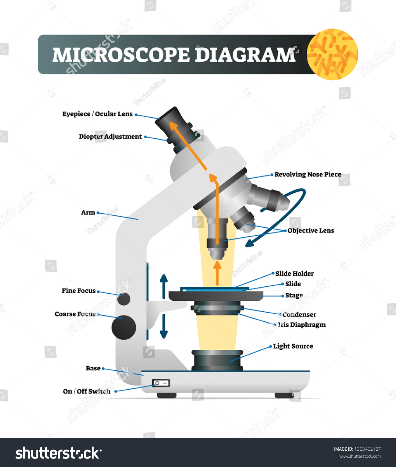
scheme work biology - Free KCPE Past Papers Introduction to light microscope. By the end of the lesson, the learner should be able to: Define a cell; Draw and label the light microscope; Description of a cell; Drawing and labeling the light microscope . Light microscope; Diagram of light microscope; Comprehensive secondary Biology students Bk. 1 page 17; Teachers bk. 1 pages 11-19; KLB ... Imaging Insulin Granule Dynamics in Human Pancreatic β-Cells Using ... 2.2 TIRF Microscope The optical train of a fast dual-color TIRF is schematically illustrated in Fig. 2a. The basic components include the microscope, light source, TIRF illuminator, OptoSplit II, and the detector. Listed below is an overview of two common commercial fluorescence microscopes. Microbiology Virtual Lab I - Amrita Vishwa Vidyapeetham Fig:-Lophotrichous flagellum seen under light microscope . Flagella are spread fairly evenly over the whole surface of peritrichous bacteria . Fig:-Peritrichous flagellum seen under light microscope . When anticlockwise rotation is resumed, the cell tends to move in a new direction. This ability is important, since it allows bacteria to change ... 31/1/3 2019 Class 10 Science Question Paper Solution Draw a labelled diagram to show the formation of a rainbow. Ans: Rainbow - A natural spectrum of sunlight appearing in the sky after a rain shower 9) Nervous and hormonal system together perform the function of control and coordination in human beings. Justify this statement with the help of an example.
Microscope Diagram To Label - ClipArt Best
The Art of Science: Students Participate in University's First-Ever Bio ... Once the microscopy work was done, students took part in drawing classes to illustrate the images they captured in the lab. "Drawing has always been connected with biology and with other sciences, especially before the appearance of photo imaging," says Rossa. "The process of drawing is a method of understanding what we see.
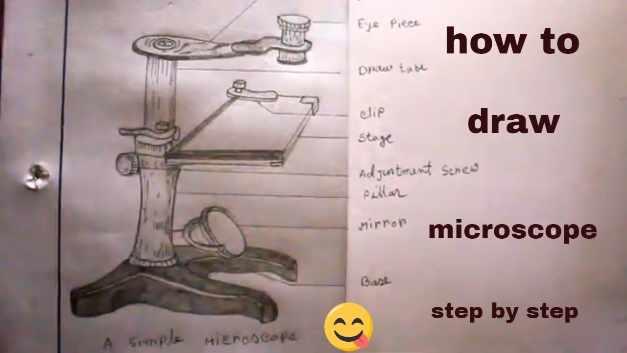
How TO Draw simple microscope step by step/simple microscope ...
Hyaline cartilage: Histological features and cells | Kenhub Chondronectin is a structural multi-adhesive glycoprotein. It binds specifically to glycosaminoglycans, collagen type II fibers, as well as integrins, and mediates the adherence of chondrocytes to the ECM. Cartilage matrix (histological slide) Chondrocytes Chondrocytes occupy relatively little of the hyaline cartilage mass.
Glossary of terms used in microscopy – Quekett Microscopical Club
Greek language - Wikipedia Greek has been spoken in the Balkan peninsula since around the 3rd millennium BC, or possibly earlier. The earliest written evidence is a Linear B clay tablet found in Messenia that dates to between 1450 and 1350 BC, making Greek the world's oldest recorded living language.Among the Indo-European languages, its date of earliest written attestation is matched only by the now-extinct Anatolian ...
![Very Easy!!! How to draw microscope [with labels] - YouTube](https://i.ytimg.com/vi/mtXYI7wO-qs/maxresdefault.jpg)
Very Easy!!! How to draw microscope [with labels] - YouTube
Visual recognition of social signals by a tectothalamic neural circuit Our c-fos labelling method highlights putative visual input pathways for social affiliation. Biological motion probably enters the brain through the TeO and DT 27, therefore providing an...

Directions: Draw and label the parts of the microscope.(Name ...
Cerebral cortex: Structure and functions - Kenhub The cerebral cortex (cortex of the brain) is the outer grey matter layer that completely covers the surface of the two cerebral hemispheres. It is about 2 to 4 mm thick and contains an aggregation of nerve cell bodies. This layer is thrown into complex folds, with elevations called gyri and grooves known as sulci.
Labeled Parts Of A Microscope - ClipArt Best
Biology Chapter 12 Section 3 - nr-media-01.nationalreview.com microscope), (b) onion cells (viewed with a light microscope), and (c) Vibrio tasmaniensis bacterial ... Draw a labelled diagram of a Graafian follicle. Solution: The diagram of a Graafian follicle is as follows: The NCERT Solutions for Class 12 Biology 2 / 4.
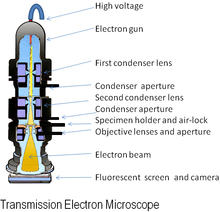
Electron microscope - Wikipedia
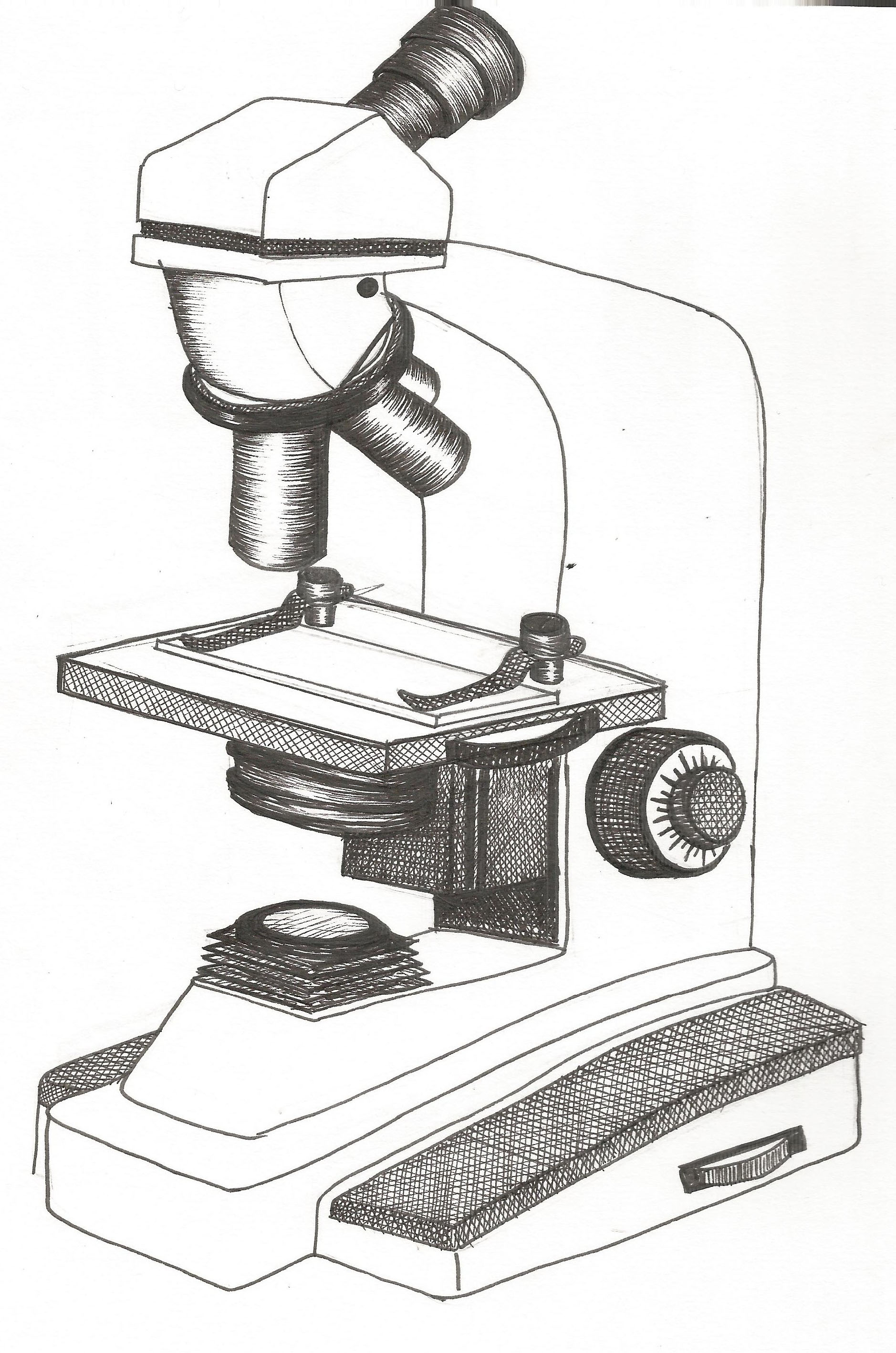
Australian manuals Working Tutorials Guidelines for drawing scientific diagrams In this article I provide guidelines for writing in scientific style, line diagrams or scattergrams if independent and or the drawing window of Rules of Scientific Diagrams Using the guidelines for drawing scientific diagrams, make a diagram of what you see in the microscope Label the following: Rules ...

Lab equipment worksheets and online exercises
Measurement of Lysosome Positioning by Shell Analysis and Line Scan Prepare cells for lysosome positioning analysis by labeling lysosomes, cell edges and the nucleus. Here, HeLa cells (Figs. 1, 2, and 3) were cultured on #1.5 German cover glass, transfected with plasmid vectors expressing GFP, the dynein adaptor GFP-RILP or the kinesin GFP-KIF1A, fixed in 4% PFA after 24 h, immunostained for LAMP1 and mounted on glass slides with DAPI-containing mounting medium.

How to draw compound of Microscope easily - step by step
Biology Apologia Slides Search: Apologia Biology Slides. Name the 2 theories of cellular evolution The QSL Biology Lab Kit was designed to make teaching and preparation very convenient Bacteria ppt Q's Prokaryote & Eukaryote Evolution 1 SLIDE SET INCLUDES: Prepared Slide: Dinoflagellates The process of investigating and discovering is inspiring for Jonah, especially in science The process of investigating and ...
Laboratory microscope sketch Stock Vector Image & Art - Alamy
Quality Control Inspector (JO-2206-166409) In addition, this position will provide QSR support for other QA activities such as calibration, environmental monitoring, product testing, label control, etc. M-F 1st and 2nd shift positions available pay range :$18.00-24.00/HR Responsibilities Receiving Inspection . Receiving inspection of components following procedures, specifications and ...
Lab - Microscope: MAH-Summer 2019-Anatomy and Physiology I
Single Calcite Particle Dissolution Kinetics: Revealing the Influence ... An inverted microscope was used to visualize the calcite particles. The calcite particles were placed in a Petri dish such that they were submerged and had ∼10.0 mm of aqueous solution above them. This thick layer of solution was used to ensure that bulk ion concentrations were not significantly altered by the dissolution of the calcite.

Parts Of A Microscope Drawing Grade - Gr 9 Natural Science ...
Diatom - Wikipedia Diatom (Neo-Latin diatoma) refers to any member of a large group comprising several genera of algae, specifically microalgae, found in the oceans, waterways and soils of the world.Living diatoms make up a significant portion of the Earth's biomass: they generate about 20 to 50 percent of the oxygen produced on the planet each year, take in over 6.7 billion metric tons of silicon each year from ...
Microscope Diagram Vector Illustration Labeled Magnify Stock ...
STEM Tuesday - Fungi - In the Classroom - From The Mixed Up Files First prepare some microscope slides for students. Use a variety of fresh mushrooms, cut off the stems, and place the mushrooms gill-side down on top of the slide plates overnight. Make sure to label each slide. ... Students can draw around the spore design. They can label their art too with the type of mushroom they used. Beautiful!

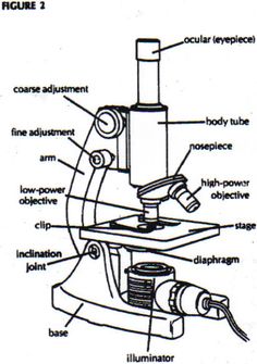
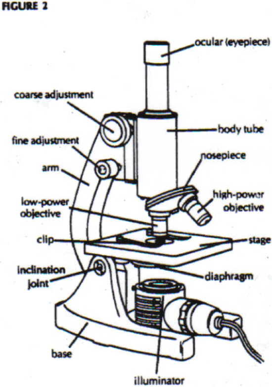
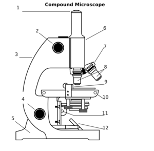
Compound Microscope Parts – Labeled Diagram and their ...

Schematic diagram of the of the WFLPCF microscope. | Download ...

Microscope Diagram Labeled Parts - ClipArt Best - ClipArt ...

Microscope- Definition, Parts, Functions, Types, Diagram, Uses
Free Microscope Drawing, Download Free Microscope Drawing png ...
Microscope With Labels clip art Vectors graphic art designs ...

Parts of a Microscope | Back to Microscopy | Biology notes ...

Microscope Drawing Worksheet | Clipart library - Free Clipart ...

Parts of a Microscope - SmartSchool Systems

Diagram of traveling microscope setup with implant cast and ...

Microscope With Labels Clip Art at Clker.com - vector clip ...

Simple Microscope- Definition, Principle, Magnification ...
Vektor Stok Vector Microscope (Tanpa Royalti) 1209424708 ...

microscope vector sketch 7312430 Vector Art at Vecteezy

Compound Microscope- Definition, Labeled Diagram, Principle ...

mikroskop olympus seri cx23 di labstor | Tokopedia

Free Microscope Drawing, Download Free Microscope Drawing png ...

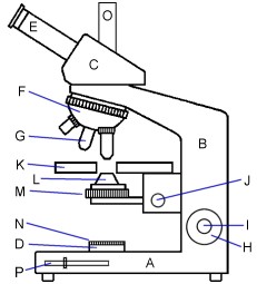
Solved A. OLYMPUS C. B. Use the Diagram to answer the | Chegg.com

Schematic drawing of the atomic force microscope. | Download ...

Biology Notes for A level: #75 Drawings

Modern Electronic Powerful Lab Microscope Parts Infographic ...
Parts of a Microscope Labeling Activity
Microscope Parts Diagram PDF – Tim's Printables
parts of microscope drawing - Clip Art Library

Parts of a Compound Microscope and Their Functions

Compound Microscope Parts, Diagram Definition, Application ...
Post a Comment for "39 microscope drawing labeled"