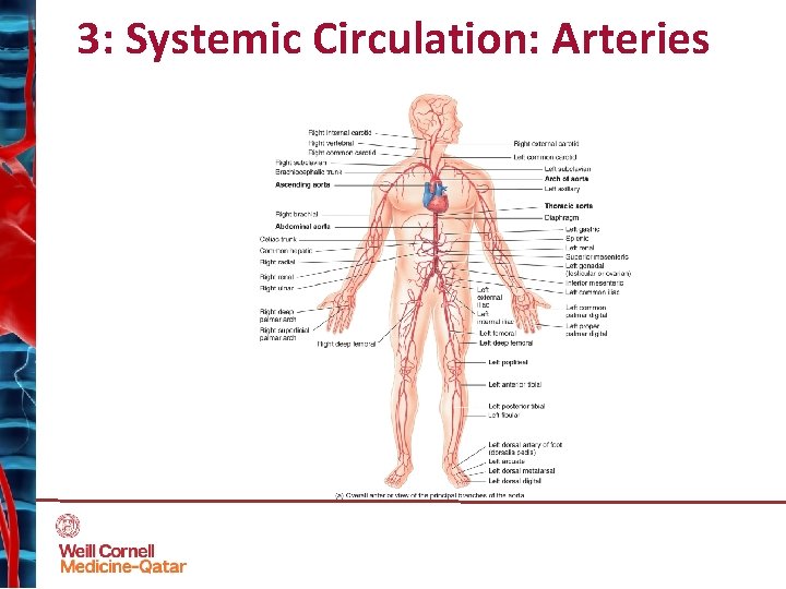
45 label the major systemic arteries
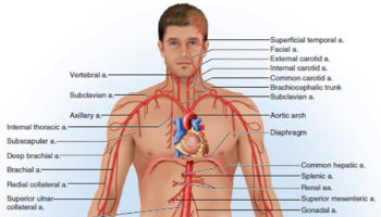
onamurazik.blogspot.com › 2022 › 04Label The Major Systemic Arteries - Cardiovascular System ... Apr 01, 2022 · A4 figure 47.17 label the major systemic arteries. Aorta (abdominal and thoracic), carotid arteries (internal and external), subclavian, axillary, . Create a flow chart showing the major systemic arteries through which blood travels from the aorta and its major branches, to the most significant arteries . › what_are_the_major_arteriesMajor Arteries of the Body: The Aorta, Head, Neck & Torso Aug 05, 2021 · The major arteries in the body are: The aorta. The largest artery in the body, which connects directly to the left ventricle of the heart. It begins the systemic division of the arterial system, which includes all the arteries that deliver blood to all the tissues in the body other than the lungs. Arteries of the head and neck (carotids)
Digi Postpaid 38 - Digi å…¨æ-°postpaid Plan 9gb 上ç½'æ•°æ ® Unlimited ... Label The Major Systemic Arteries - Cardiovascular System Health Jade Create a flow chart showing the major systemic arteries thr… Layan Drama Titian Cinta / Senarai Pelakon Titian Cinta Cerita Budak Sepet Layan drama · sinopsis titian cinta. This is titian cinta e…
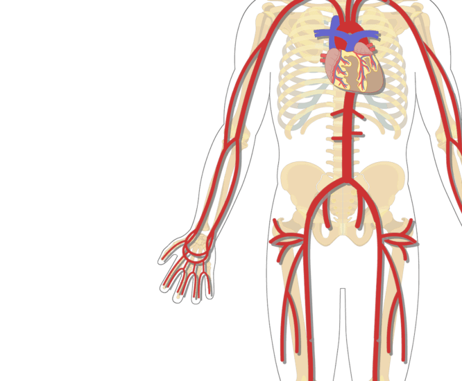
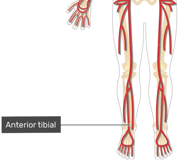
Label the major systemic arteries
What are the main pathologic findings of hypertension (high blood ... WhatsApp. Answer. The main pathologic findings are in the heart, which shows an increase in mass caused principally by left ventricular hypertrophy. Histologically, the individual myocytes are ... Atrial Septal Defect: Pathophysiology, Diagnosis, and Treatment - Medscape Larger defects are associated with substantial shunting, which may lead to volume overload of the right atrium, right ventricle, and pulmonary arteries. The magnitude of left-to-right shunting depends on the size of the ASD, the relative compliance of the 2 ventricles, and the pulmonary and systemic vascular resistance. Peripheral Artery Disease. Part 1: Clinical Evaluation and ... - Medscape One candidate biomarker for PAD that emerged from a proteomic screening is beta 2 microglobulin, a component of the major histocompatibility complex class I proteins involved in the regulation of immune and inflammatory pathways. Cooke and Wilson reported that this marker was elevated in patients with PAD and correlated with the severity of ...
Label the major systemic arteries. The Cerebrospinal Venous System: Anatomy, Physiology, and ... - Medscape Groen [29] The fundamental feature that distinguishes the CSVS from the systemic (caval) venous system is the lack of venous valves. In 1940, Batson [2] demonstrated that the VVS was angiographically linked to the cranial venous system, and that retrograde flow from the VVS into the brain was possible because of the lack of venous valves. Kedai Cermin Meja Makan - Kedai Jual Sarung Kerusi Makan Kerusireview Kedai Cermin Meja Makan - Kedai Jual Sarung Kerusi Makan Kerusireview Migraine - Wikipedia A migraine (UK: / ˈ m iː ɡ r eɪ n /, US: / ˈ m aɪ-/) is a neurological disorder characterized by recurrent headaches that are moderate to severe. Typically, episodes affect one side of the head, are pulsating in nature, and last from a few hours to three days. Associated symptoms may include nausea, vomiting, and sensitivity to light, sound, or smell. The pain is generally made worse by ... › blood-vessels › major-arteriesMajor Systemic Arteries | GetBodySmart Nov 29, 2017 · Review the major systemic veins of the body including the veins of the neck, arm, forearm, abdomen, pelvis, thigh, and leg in this interactive tutorial. Aorta Anatomy An illustrated and interactive tutorial covering the anatomy of the aorta, the largest artery of the human body.
› homework-help › questions-andSolved Jonso14 Label the major systemic arteries of ... - Chegg The labeled figure of major arteries is attached below- Arteries are the blood vessels that carry oxygenated blood to every tissue and organ of the body from the heart. Carotid artery- The two carotid arteries carry the oxygenated blood to the head a … Organs of Circulatory System and Their Functions - New Health Advisor 1. The Heart Located slightly to the left of the middle of your chest, the heart is made of strong muscle tissue and is protected by your rib cage. Even though it is no larger than the size of your fist, it plays a vitally important role in your body. It consists of four hollow chambers - two ventricles and two atria. Diagram of Human Heart and Blood Circulation in It There are three arteries of the heart, including pulmonary artery, aorta, and coronary arteries. Pulmonary Circulation Route and Process | New Health Advisor The pulmonary arteries transport blood low in oxygen from heart's right side to the two lungs. The pulmonary veins, on the other hand, transport oxygen rich blood to heart's left side. Exchange of Substances in Pulmonary Capillaries The circulatory system in humans as well as in other mammals is basically a closed circuit.
65 Fun Facts About The Circulatory System: What Do You Need To ... - Kidadl The circulatory system consists of five main parts, blood, the heart, blood vessels, arteries, capillaries and veins. 2. Five different types of blood vessels make up the human circulatory system, and they are the arteries, arterioles, capillaries, venules and veins. 3. Blood travels away from the left side of the heart. 4. Organ of Zuckerkandl | Radiology Reference Article - Radiopaedia The organ of Zuckerkandl comprises of a small mass of chromaffin cells derived from neural crest located along the aorta, beginning cranial to the superior mesenteric artery or renal arteries and extending to the level of the aortic bifurcation or just beyond. The highest concentration is typically seen at the origin of the inferior mesenteric ... quizlet.com › 637928144 › chapter-13-ap-circulatoryChapter 13 A&P circulatoryII-Heart and blood - Quizlet Correctly label the following major systemic arteries. Correctly label the following veins of the head and neck. Other sets by this creator. Chapter 16 continued. 15 ... Circulatory system - Wikipedia The circulatory system includes the heart, blood vessels, and blood. The cardiovascular system in all vertebrates, consists of the heart and blood vessels. The circulatory system is further divided into two major circuits - a pulmonary circulation, and a systemic circulation. The pulmonary circulation is a circuit loop from the right heart taking deoxygenated blood to the lungs where it is ...
Cardiovascular System
Definition and types of connective tissue - Kenhub The primary cell of connective tissue is the fibroblast.Its function is to produce and maintain the ECM of connective tissue. Besides fibroblasts, several other cell types are present. These are the cells of the immune system (macrophages, lymphocytes and mast cells) and adipocytes. Specialised connective tissue contains specialised cells, for example cartilage contains chondrocytes and bone ...

Nikaya Woranansit (woranansit) - Profile | Pinterest
› homework-help › questions-andSolved Label the major systemic arteries by clicking and ... Label the major systemic arteries by clicking and dragging the labels to the correct location. 7 Ulnar a. Aorta Brachial a. Femoral a Common carotid a. External iliac a Common iliac Popliteal a. Dorsalis pedis a. Radial a Brachiocephalic trunk Axillary a Subclavian a

veins+capillaries+arteries | the aorta and distributed to ...
Baju Bowling Sublimation : 30 95 - Ona Murazik Adding trendy new items to the roster is easy with this wide selection of men's . Alogo sportswear polo shirt sport jersey training gear dry fit baju sejuk lelaki unisex sports sukan.
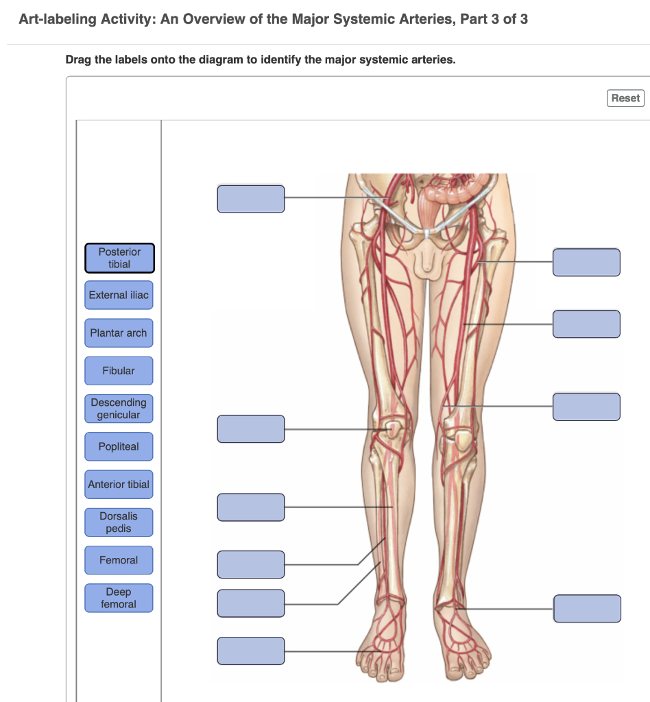
Solved Art-labeling Activity: An Overview of the Major ...
What are the AHA/ASA guidelines on thrombolytic therapy ... - Medscape Safety and efficacy of desmoteplase given 3-9 h after ischaemic stroke in patients with occlusion or high-grade stenosis in major cerebral arteries (DIAS-3): a double-blind, randomised, placebo ...

Circulatory Pathways | Anatomy and Physiology II
Path of Blood Through the Heart - New Health Advisor The pulmonary arteries distribute blood to the lungs. Blood circulates in the lungs, where oxygen (O2) is added to the bloodand carbon dioxide (CO2) is removed. Blood returns to the heart through the pulmonary veins, which go into the left chambers. 4. Left Atrium
Major Systemic Arteries | GetBodySmart
What Are Arteries, Veins And Capillaries? - Science ABC The arteries are blood vessels that carry oxygenated blood from the heart to all the organs. The veins are meant to transport deoxygenated or blood devoid of oxygen from the organs to the heart so that it can gain oxygen again. Capillaries are the tiniest blood vessels of the body and serve as the transition link between arteries and veins.

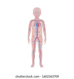
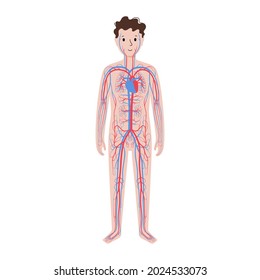
Full body front view of the male figure with the skeleton ...
Lung - Wikipedia The lungs are the primary organs of the respiratory system in humans and most animals including a few fish, and some snails.In mammals and most other vertebrates, two lungs are located near the backbone on either side of the heart.Their function in the respiratory system is to extract oxygen from the air and transfer it into the bloodstream, and to release carbon dioxide from the bloodstream ...

Johny's A & P Blood Circulation
Heart - Wikipedia The wall of the heart is made up of three layers: epicardium, myocardium, and endocardium. [7] The heart pumps blood with a rhythm determined by a group of pacemaker cells in the sinoatrial node. These generate a current that causes the heart to contract, traveling through the atrioventricular node and along the conduction system of the heart.

Potential health benefits of functional components | Download ...
Arterial Aging -- Hemodynamic Changes and Therapeutic Options Over the past 50 years, one of the major findings from studies of cardiovascular disease has been the proven effectiveness of antihypertensive therapy. These treatments are beneficial for the prevention of stroke, heart failure, renal insufficiency and, to a lesser extent, coronary artery disease (CAD).

AHCDW16Notes13.pdf - 13 Award 1.00 point Problems Adjust ...
All You Need to Know about Chronic Microvascular Ischemic Disease It is the result of blockage to the small blood vessels in the brain. This happens over time due to changes in the blood vessels or blood clots. Ischemic changes are areas in the brain tissue that have died from lack of blood flow. Microvascular changes are so small that the disease may never cause any symptoms and the disease is usually found ...

The Major Systemic Veins Part 2 of 3 Diagram | Quizlet
Circulatory System Diagram | New Health Advisor Arteries These blood vessels are responsible for carrying the blood from heart to different parts of body. Almost all the arteries carry oxygenated blood. Arteries have thick wallsor in other words arterial walls have a thicker muscle coat. The pressure of blood flow is highest as soon as the blood enters in aorta from the left ventricle.

AHCDW16Notes15.pdf - 15. Award: 1.00 point Problems? Adjust ...
12.1: Introduction to the Cardiovascular System - Blood Vessels and ... Label the major blood vessels of the pulmonary and systemic circulations Identify and describe the hepatic portal system Describe the development of blood vessels and fetal circulation Compare fetal circulation to that of an individual after birth

Principal Systemic Arteries The Arterial System Carries ...
Coronary artery disease - Wikipedia Coronary artery disease ( CAD ), also called coronary heart disease ( CHD ), ischemic heart disease ( IHD ), [13] myocardial ischemia, [14] or simply heart disease, involves the reduction of blood flow to the heart muscle due to build-up of atherosclerotic plaque in the arteries of the heart.
Untitled
What is the role of the anterior and middle cerebral arteries in the ... The middle cerebral artery (MCA) supplies the lateral portions of the frontal and parietal lobes, as well as the anterior and lateral portions of the temporal lobes, and gives rise to perforating...

Circulatory Pathways | Anatomy and Physiology II
Systolic vs. Diastolic Blood Pressure - Verywell Health Systolic blood pressure is the pressure exerted when the heart beats and blood is ejected into the arteries. In a blood pressure measurement written as a fraction, the systolic blood pressure is the top number. Normal systolic blood pressure is 120 mmHg or lower. 7 What is diastolic blood pressure?
Major Systemic Arteries and Veins of the Body
Common Diseases of the Circulatory System | MD-Health.com Arteriosclerosis is a common disease of the circulatory system caused by the buildup of fat, cholesterol, or other substance in the artery wall. Deposits in the artery cause the vessel to stiffen and narrow. Diabetes, high cholesterol, smoking, and high blood pressure can result in stiff arteries that restrict blood flow through the heart.

Cardiovascular System
Blood vessels and nerves of the eye: Anatomy - Kenhub The veins that drain into it are named as the arteries of the region (medial palpebral, lacrimal, anterior ethmoidal, inferior ophthalmic, central retinal and muscular). The inferior ophthalmic vein runs over the surface of the inferior rectus muscle, and drain to the cavernous sinus or the superior ophthalmic vein.

Major Veins & Arteries of Systemic Circulation Quiz
Systemic Sclerosis: An Integrated Approach to Understanding ... Off-Label This activity contains discussion of unlabeled and non-FDA-approved uses of commercial products. ... which is the major cause of chronic renal problems. [ CLOSE WINDOW] Slide 14. Endothelin Receptor Blockade With Bosentan. Vascular Pathology at Multiple Sites: The Kidney This is a common vascular pathway, and we viewed this picture of ...
Major arteries, veins and nerves of the body: Anatomy | Kenhub
Peripheral Artery Disease. Part 1: Clinical Evaluation and ... - Medscape One candidate biomarker for PAD that emerged from a proteomic screening is beta 2 microglobulin, a component of the major histocompatibility complex class I proteins involved in the regulation of immune and inflammatory pathways. Cooke and Wilson reported that this marker was elevated in patients with PAD and correlated with the severity of ...

Blood supply Stock Photos and Images | agefotostock
Atrial Septal Defect: Pathophysiology, Diagnosis, and Treatment - Medscape Larger defects are associated with substantial shunting, which may lead to volume overload of the right atrium, right ventricle, and pulmonary arteries. The magnitude of left-to-right shunting depends on the size of the ASD, the relative compliance of the 2 ventricles, and the pulmonary and systemic vascular resistance.
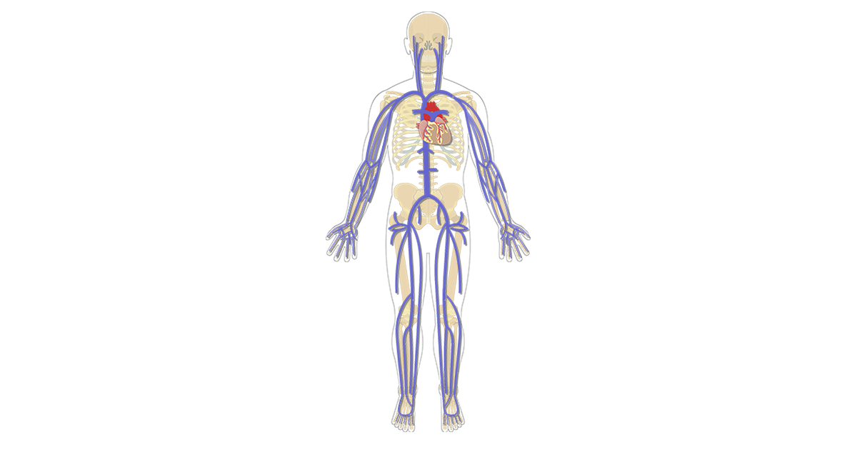
The Major Systemic Veins | GetBodySmart
What are the main pathologic findings of hypertension (high blood ... WhatsApp. Answer. The main pathologic findings are in the heart, which shows an increase in mass caused principally by left ventricular hypertrophy. Histologically, the individual myocytes are ...
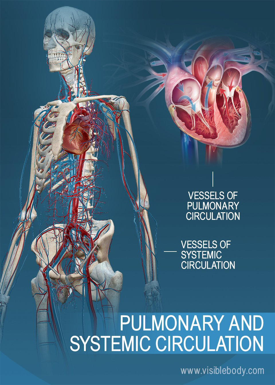
Pulmonary & Systemic Circulation | Circulatory Anatomy
Articles - Page 466 of 505 - Health Jade

Circulatory Pathways | Anatomy and Physiology II

Major Systemic Blood Vessels Flashcards | Quizlet
NVCC Bio 212
![Cardiovascular system[1]](https://image.slidesharecdn.com/aa2crr3qquena9c7vv5a-signature-460517c25b85fc4e63c8080c3e27df73c8dfae9e0c6544cc7ea6d9e8b5e79cc7-poli-180213064029/85/cardiovascular-system1-16-320.jpg?cb=1518504621)
Cardiovascular system[1]

Correctly label the following major systemic veins. Cephalic ...

Chapter 12 The Cardiovascular System - ppt video online download
Human Development Structure Fall 2019 Blood Vessels 1

Blood vessel LABS Flashcards | Quizlet

Major Systemic Arteries Diagram | Quizlet
Vector Isolated Illustration Human Arterial Venous Stock ...

On vasa vasorum: A history of advances in understanding the ...

Pre-existing Vascular Issues - TrialExhibits Inc.

Major arteries Quiz - By anatomygirl1995

AHCDW16Notes14.pdf - 14. Award: 1.00 point Problems? Adjust ...
Veins arteries - Teaching resources
Cardiovascular System

mucles Anterior Muscles Origin on pelvis or vertebral
Major Systemic Arteries | GetBodySmart
390 Carotid body Images, Stock Photos & Vectors | Shutterstock
Veins arteries - Teaching resources

Major Veins and Arteries | Biology - Quizizz

Blood Vessels Types of Blood Vessels Arteries vessels
Post a Comment for "45 label the major systemic arteries"