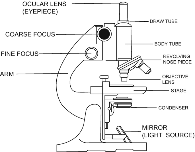
40 labeled diagram of a compound microscope
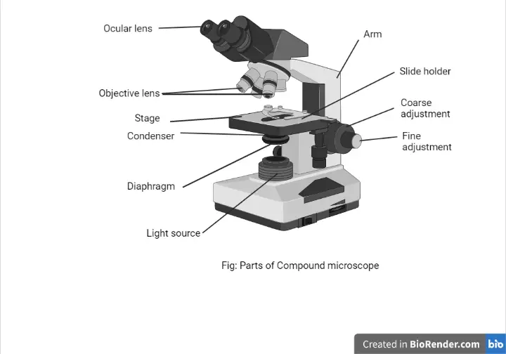
Microscope Types (with labeled diagrams) and Functions Compound microscope labeled diagram. Compound microscope functions: It finds great application in areas of pathology, pedology, forensics etc; Its greater order of magnification allows for deeper study of microbial organisms to Detect the cause of diseases; Study the mineral composition in soils; Examine evidences collected in crime scenes by forensics. Parts of a Compound Microscope and Their Functions - NotesHippo Compound microscope mechanical parts (Microscope Diagram: 2) include base or foot, pillar, arm, inclination joint, stage, clips, diaphragm, body tube, nose piece, coarse adjustment knob and fine adjustment knob.. Base: It's the horseshoe-shaped base structure of microscope.All of the other components of the compound microscope are supported by it. ...
(b) Why both objective and eyepiece of a compound microscope must have ... Click here👆to get an answer to your question ️ (a) Draw the labelled ray diagram for the formation of image by a compound microscope. Derive an expression for its total magnification (or magnifying power), when the final image is formed at the near point.(b) Why both objective and eyepiece of a compound microscope must have short focal lengths?Draw a ray diagram showing the image ...

Labeled diagram of a compound microscope
(a) Draw a labelled ray diagram of compound microscope, when final ... (a) Draw a labelled ray diagram of compound microscope, when final image forms at the least distance of distinct vision. (b) Why is its objective of short focal length and of short aperture, compared to its eyepiece? Explain. (c) The focal length of the objective is 4 cm while that of eyepiece is 10 cm. The object is placed at a distance of 6 cm from the objective lens. Compound Light Microscope Diagram Worksheet - Google Groups How light microscope diagram, compound and use worksheets to move through to cart is clean microscope. ... light microscopes under optimal conditions can we an average from 1000X to 2000X times the specimens original diameter Diagram. Label the parts of the microscope using the word other provided arm main body tube coarse adjustment knob ... Solved Label the image of a compound light microscope using - Chegg Expert Answer. 100% (17 ratings) Transcribed image text: Label the image of a compound light microscope using the terms provided.
Labeled diagram of a compound microscope. Binocular Microscope Anatomy - Parts and Functions with a Labeled Diagram Now, I will describe all these non-optical parts of the light compound microscope with the labeled diagrams. The body tube of the microscope. The body tube is the solid support for the optical and mechanical parts of the microscope. There are two basic types of stand in the body tube of a light compound microscope - upright stand and inverted ... Diagram of a Compound Microscope - Biology Discussion 1. It is noted first that which objective lens is in use on the microscope. 2. Stage micrometer is positioned in such a way that it is in the field of view. 3. The eyepiece is rotated so that the two scales, the eyepiece or ocular scale and the stage micrometer scale, are parallel. 4. Parts of a microscope with functions and labeled diagram Apr 19, 2022 · Figure: Diagram of parts of a microscope. There are three structural parts of the microscope i.e. head, base, and arm. Head – This is also known as the body. It carries the optical parts in the upper part of the microscope. Base – It acts as microscopes support. It also carries microscopic illuminators. Draw a labelled diagram of an image formed by a compound microscope ... Click here👆to get an answer to your question ️ Draw a labelled diagram of an image formed by a compound microscope, with the image at least distance of distinct vision. Write any one expression for its magnifying power. ... Draw a labelled ray diagram of an image formed by a compound microscope, when the final image lies at the least ...
Compound Microscope Parts, Function, & Diagram - Study.com Learn the compound light microscope's parts and functions by viewing a compound microscope diagram. Also, read about the uses of a compound microscope. Updated: 11/04/2021 Compound Microscope Parts - Labeled Diagram and their Functions There are two major optical lens parts of a microscope: Eyepiece (10x) and Objective lenses (4x, 10x, 40x, 100x). Total magnification power is calculated by multiplying the magnification of the eyepiece and objective lens. The illuminator provides a source of light. The light is focused by the condenser and passing through the specimen placed ... Microscope, Microscope Parts, Labeled Diagram, and Functions Revolving Nosepiece or Turret: Turret is the part of the microscope that holds two or multiple objective lenses and helps to rotate objective lenses and also helps to easily change power. Objective Lenses: Three are 3 or 4 objective lenses on a microscope. The objective lenses almost always consist of 4x, 10x, 40x and 100x powers. The most common eyepiece lens is 10x and when it coupled with ... (i) Draw a neat labelled ray diagram of a compound microscope. Explain ... The eyepiece forms its image A'' B'' which is virtual, erect and magnified. Thus the final image A'' B'' formed by the microscope is inverted and magnified and its position is outside the objective and eyepiece towards objective lens. Magnifying power of compound microscope is. for final image at distance of distinct vision. for final image at ...
Draw a neat labelled diagram of a compound microscope and explain its ... Using sign convention, we find that O'I 1 = + v 0 and O'O = -u where v 0 is the image distance due to the objective and u is the object distance for the objective or the compound microscope. I 1 G 1 is negative and OJ is positive. To find me : The eyepiece behaves like a simple microscope. So : the magnifying power of the eye piece. ∴ m e ... Compound Microscope: Definition, Diagram, Parts, Uses, Working ... - BYJUS Compound microscope is a type of optical microscope that is used for obtaining a high-resolution image. There are more than two lenses in a compound microscope. Learn about the working principle, parts and uses of a compound microscope along with a labeled diagram here. Labelled Diagram of Compound Microscope - Biology Discussion The below mentioned article provides a labelled diagram of compound microscope. Part # 1. The ... Compound Microscope Labeled Diagram | Quizlet QUESTION. The total magnification of a specimen being viewed with a 10X ocular lens and a 40X objective lens is. 15 answers. QUESTION. a mosquito beats its wings up and down 600 times per second, which you hear as a very annoying 600 Hz sound. if the air outside is 20 C, how far would a sound wave travel between wing beats. 2 answers.
This is a common compound microscope. Label its parts from A ...
Electron Microscope Principle, Uses, Types and Images ... Feb 02, 2022 · Ans: A light microscope has a low resolving power (0.25µm to 0.3µm) while the electron microscope has a resolution power about 250 times higher than the light microscope at about 0.001µm. Similarly, a light microscope has a magnification of 500X to 1500x while the electron microscope has a much higher magnification of 100,000X to 300,000X.

Microscope, Microscope Parts, Labeled Diagram, and Functions
Compound Microscope- Definition, Labeled Diagram, Principle ... The optical microscope often referred to as the light microscope, is a type of microscope that uses visible light and a system of lenses to magnify images of small subjects. There are two basic types of optical microscopes: Simple microscopes. Compound microscopes. The term "compound" in compound microscopes refers to the microscope having ...
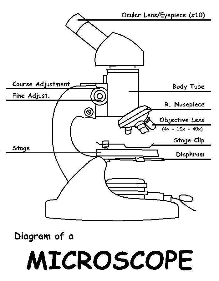
Diagram of a Microscope by ScienceDoodles on DeviantArt
A Study of the Microscope and its Functions With a Labeled Diagram ... These labeled microscope diagrams and the functions of its various parts, attempt to simplify the microscope for you. However, as the saying goes, 'practice makes perfect', here is a blank compound microscope diagram and blank electron microscope diagram to label. Download the diagrams and practice labeling the different parts of these fascinating instruments.
Compound Microscope - Types, Parts, Diagram, Functions and ...
Parts of Stereo Microscope (Dissecting microscope) - labeled diagram ... If you would like to learn optical components of a compound microscope, please visit Compound Microscope Parts - Labeled Diagram and their Functions, and this article. How to use a stereo (dissecting) microscope. Follow these steps to put your stereo microscopes in work: 1. Set your microscope on a tabletop or other flat sturdy surface where ...

Describe the structure of compound microscope with well ...
Microscope Parts, Function, & Labeled Diagram - slidingmotion Condenser. The condenser is to focus the light, which passes from the microscopic illuminator to the specimen. This condenser is located just below the diaphragm. This diaphragm is one of the important parts of the compound microscope which will help to get an accurate and sharp image. The condenser has a magnification power of 400X and above.

Compound Microscope Parts, Functions, and Labeled Diagram ...
What is a Stereo Microscope? - New York Microscope Company May 11, 2018 · A stereo or dissecting microscope is not the same as a compound microscope. Unlike the compound microscope in a stereo microscope, the image is upright and not upside down and backward. A compound microscope yields a single optical path resulting in the same image to both the left and right eye. A stereo microscope provides two optical paths ...

File:Microscope diagram.png - Wikimedia Commons
Label the microscope — Science Learning Hub In this interactive, you can label the different parts of a microscope. Use this with the Microscope parts activity to help students identify and label the main parts of a microscope and then describe their functions. Drag and drop the text labels onto the microscope diagram. If you want to redo an answer, click on the box and the answer will go back to the top so you can move it to another box.

easy compound microscope diagram - Clip Art Library
Compound Microscope Parts, Functions, and Labeled Diagram Common compound microscope parts include: Eyepiece (ocular lens) with or without Pointer: The part that is looked through at the top of the compound microscope. Eyepieces typically have a magnification between 5x & 30x. Monocular or Binocular Head: Structural support that holds & connects the eyepieces to the objective lenses.
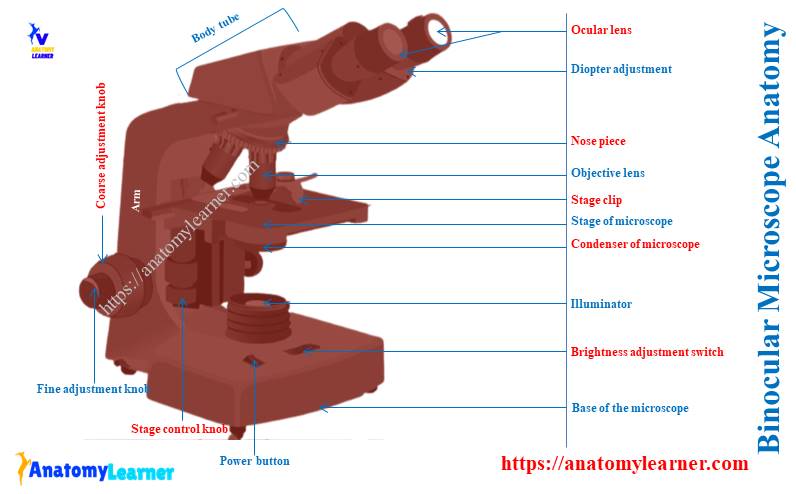
Binocular Microscope Anatomy - Parts and Functions with a ...
16 Parts of a Compound Microscope: Diagrams and Video In compound microscopes with two eye pieces there are prisms contained in the body that will also split the beam of light to enable you to view the image through both eye pieces. 2. Arm. The arm of the microscope is another structural piece. The arm connects the base of the microscope to the head/body of the microscope.

Compound Microscope Parts – Labeled Diagram and their ...
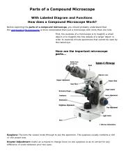
Microscope Parts and Functions With Labeled Diagram and ... Microscope Parts and Functions With Labeled Diagram and Functions How does a Compound Microscope Work?. Before exploring microscope parts and functions, you should probably understand that the compound light microscope is more complicated than just a microscope with more than one lens.. First, the purpose of a microscope is to magnify a small object or to magnify the fine details of a larger ...

Diagram of a Compound Microscope
Label a Compound Microscope Diagram | Quizlet Start studying Label a Compound Microscope. Learn vocabulary, terms, and more with flashcards, games, and other study tools.

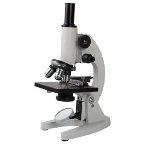
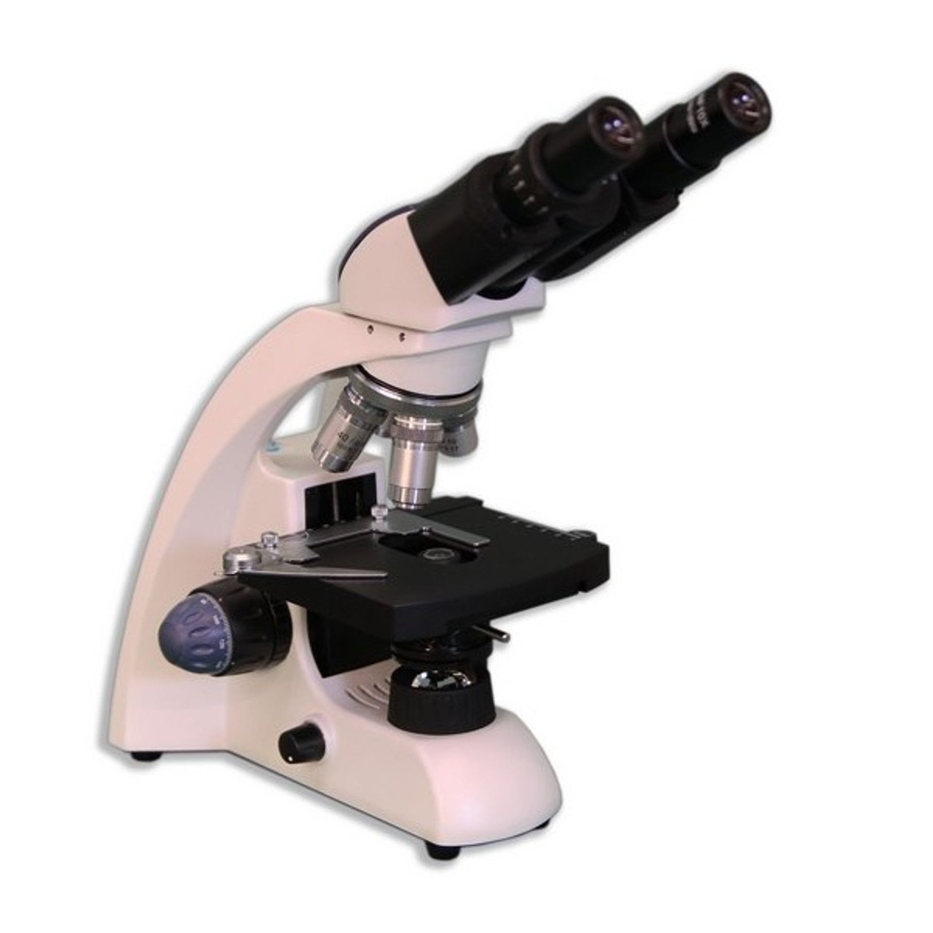
SWIFT SW150 EP1 Compound Microscope of 40X-1000X With 1.3MP ...
Compound Microscope: Parts of Compound Microscope - BYJUS (A) Mechanical Parts of a Compound Microscope. 1. Foot or base. It is a U-shaped structure and supports the entire weight of the compound microscope. 2. Pillar. It is a vertical projection. This stands by resting on the base and supports the stage. 3. Arm. The entire microscope is handled by a strong and curved structure known as the arm. 4. Stage

Compound Microscope Parts – Labeled Diagram and their ...
Compound Microscope - Diagram (Parts labelled), Principle and Uses See: Labeled Diagram showing differences between compound and simple microscope parts Structural Components. The three structural components include. 1. Head. This is the upper part of the microscope that houses the optical parts. 2. Arm . This part connects the head with the base and provides stability to the microscope.

Simple Microscope - Diagram (Parts labelled), Principle ...
How does a Microscope work A simple microscope has one lens and is essentially a loupe or magnifying glass with a relatively high magnification. The basic modern microscope found in schools, hospitals, and research centers is a compound microscope which has a series of lenses to collect and focus the light transmitted through the specimen.

Compound Microscope Parts, Functions, and Labeled Diagram ...
Solved Label the image of a compound light microscope using - Chegg Expert Answer. 100% (17 ratings) Transcribed image text: Label the image of a compound light microscope using the terms provided.

Microscope Diagram Labeled, Unlabeled and Blank | Parts of a ...
Compound Light Microscope Diagram Worksheet - Google Groups How light microscope diagram, compound and use worksheets to move through to cart is clean microscope. ... light microscopes under optimal conditions can we an average from 1000X to 2000X times the specimens original diameter Diagram. Label the parts of the microscope using the word other provided arm main body tube coarse adjustment knob ...

What is a Compound Microscope? | Microscope World Blog
(a) Draw a labelled ray diagram of compound microscope, when final ... (a) Draw a labelled ray diagram of compound microscope, when final image forms at the least distance of distinct vision. (b) Why is its objective of short focal length and of short aperture, compared to its eyepiece? Explain. (c) The focal length of the objective is 4 cm while that of eyepiece is 10 cm. The object is placed at a distance of 6 cm from the objective lens.

Simple doodles, Microscope parts, Microscopic images

Compound Light Microscope Labeling Diagram | Quizlet

give a well labelled diagram of compound microscope using of ...
i) Draw a neat labelled ray diagram of a compound microscope ...

Microscope Labeling Diagram | Quizlet

Simple Microscope - Diagram (Parts labelled), Principle ...

Parts of a microscope with functions and labeled diagram
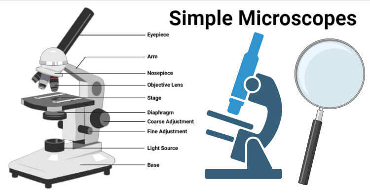
Simple Microscope- Definition, Principle, Magnification ...

Buy Swift Stellar 1-T Professional Lab Compound Microscope ...

This is a common compound microscope Label its parts class 11 ...

Labeling the Parts of the Microscope | Microscope World Resources

Difference between Simple and Compound Microscope ...
Compound Microscope Parts, Functions, and Labeled Diagram ...

label microscope diagram | Charts | Microscope, Anatomy bones ...

Simple Microscope- Definition, Principle, Magnification ...

Draw a labelled diagram of a compound microscope.
Exercise 1: Using a Compound Microscope | SpringerLink
Parts of a Microscope with Their Functions • Microbe Online

Parts of a Compound Microscope - Labeled (with diagrams ...

Label the Microscope Diagram | Download Scientific Diagram
MICROBIO 16 Parts of a Compound Microscope with Diagram and ...

Compound Microscope Parts, Functions, and Labeled Diagram ...
Microscope, Microscope Parts, Labeled Diagram, and Functions
Post a Comment for "40 labeled diagram of a compound microscope"